
Fig 1.
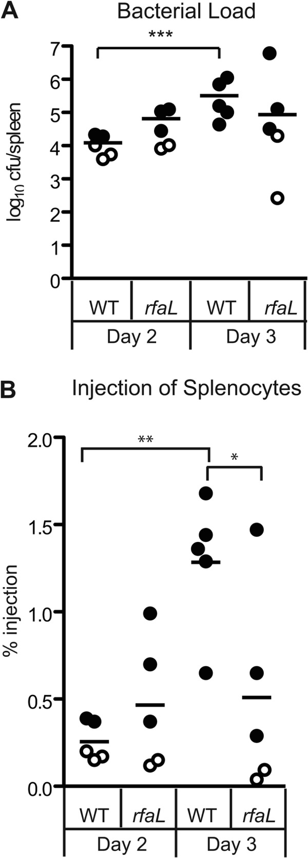
In vivo injection phenotype of the wild type and the ΔrfaL mutant. Six- to 8-week-old C57BL/6 mice were infected with the wild type or the ΔrfaL mutant carrying YopM-Bla. Spleens were harvested 3 days postinfection and homogenized. (A) Colonization of spleens by the wild type and the ΔrfaL mutant. Diluted spleen homogenates were plated in triplicate, and means are shown. The limit of detection was 102 CFU. Data were analyzed by two-tailed Student's t test (***, P < 0.001). (B) Injection of splenocytes by the wild type and the ΔrfaL mutant. The homogenized spleens were stained with CCF2-AM, flow cytometry was performed to determine the percentage of blue cells per spleen, and means are shown. Data were analyzed by one-way ANOVA and the Bonferroni post hoc test. **, P < 0.01; *, P < 0.05. Circles, YopM-Bla. Open circles correspond to samples that had only background levels of blue cells, similar to the glutathione S-transferase–Bla negative control (not shown).