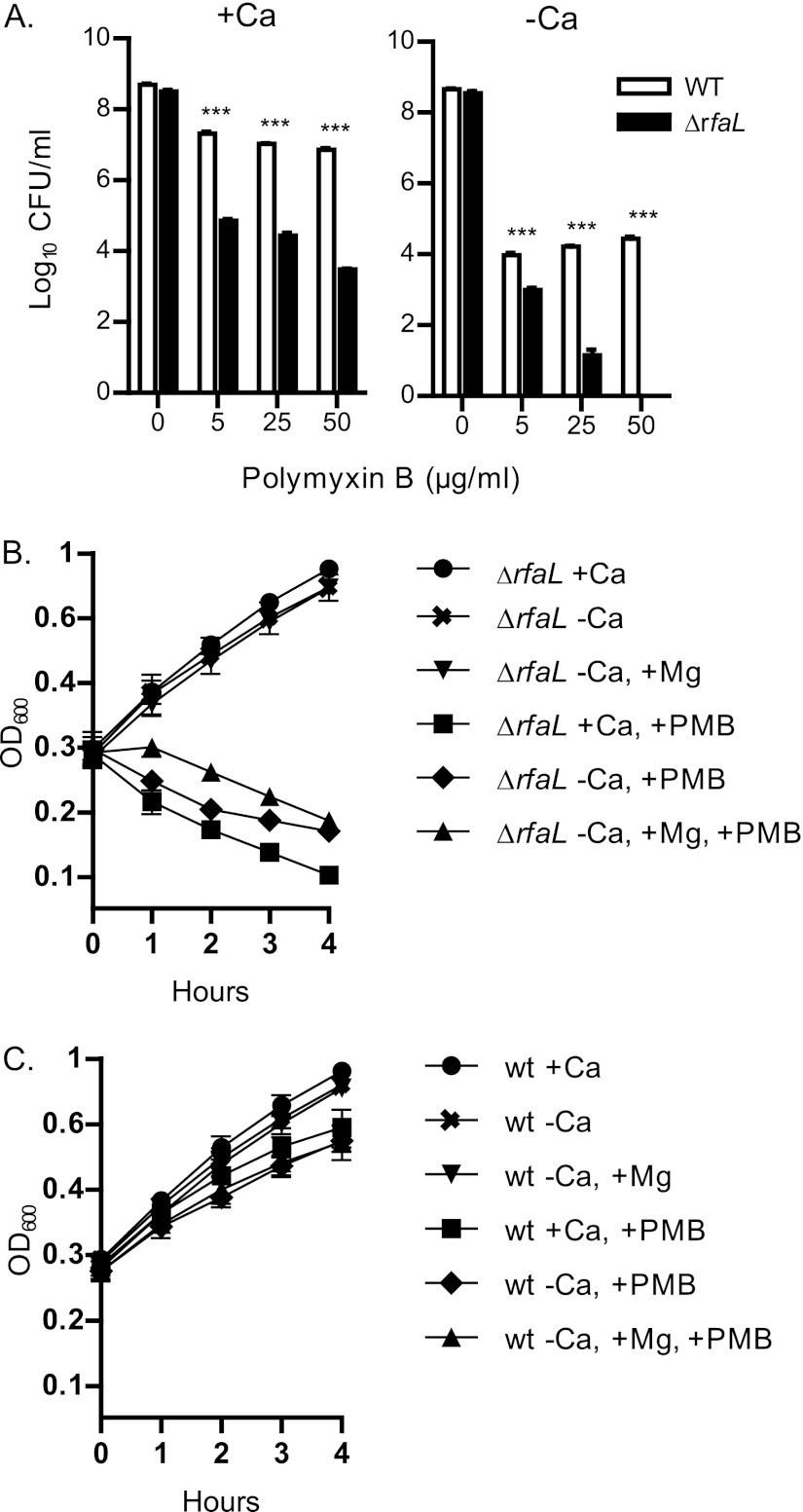
Fig 8.
Polymyxin B sensitivity assays. (A) The wild type and the ΔrfaL mutant were serially diluted and plated in triplicate onto HIA plates with or without added sodium oxalate (−Ca or +Ca) containing the indicated concentrations of polymyxin B (PMB). Plates were incubated for 2 days at 26°C. Colonies were counted, and the average log10 CFU/ml was determined. The data shown are representative of three independent trials. Significance was determined using one-way ANOVA and the Bonferroni post hoc test. ***, P < 0.001. (B and C) The wild type and the ΔrfaL mutant were diluted to an OD600 of ∼0.2 into DMEM with or without EGTA (−Ca or +Ca). Where noted, MgSO4 and/or polymyxin B was added. The cultures were incubated at 26°C, and the OD600 of each culture was taken every hour for 4 h. The experiment was performed in triplicate, and the data were pooled. Shown are the means and SEM of the combined trials.

