
Fig 3.
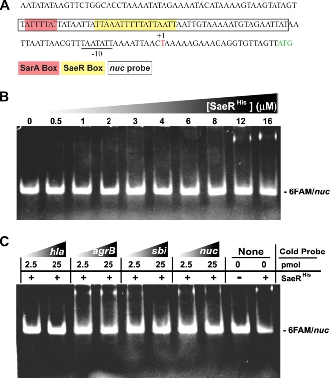
The nuc transcript start site and SaeR binding to the nuc promoter. (A) Sequence results generated by 5′ RACE with nuc-specific primers in Table 2. The nuc transcript start site (+1) and putative −10 and −35 promoter regions are underlined. The Nuc start codon is in green, and the nuc promoter probe sequence (50 bp) used for EMSA experiments is boxed. Putative SaeR (yellow) and SarA (red) boxes are shown. (B) EMSA with increasing concentrations of SaeRHis (0, 0.5, 1, 2, 3, 4, 6, 8, 12, and 16 μmol/liter), 0.5 pmol of the FAM-labeled nuc promoter probe, and 500 ng of salmon sperm DNA. (C) Binding of SaeRHis (16 μmol/liter) to the nuc promoter probe (0.5 pmol) in the presence of 2.5 or 25 pmol of the indicated unlabeled promoter and 500 ng of salmon sperm DNA.