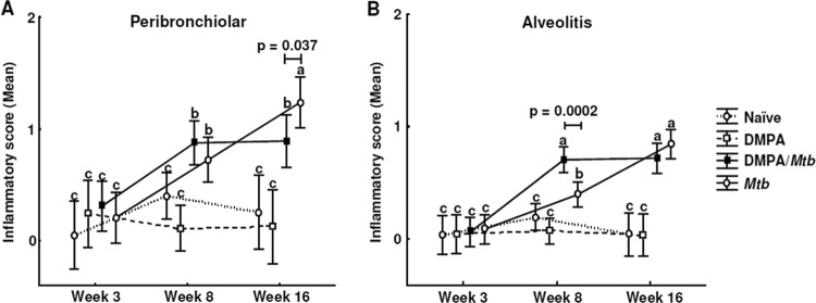
Fig 9.

DMPA alters lymphocytic infiltration in lungs of M. tuberculosis-infected BALB/c mice. Mice were injected weekly with MPA or saline and infected with a low aerosol dose of H37Rv 1 week after initiation of DMPA injections. Histopathological scoring was done on the left lung lobes in a blinded fashion in two independent experiments, and tissue was scored from 0 to 4 for the extent of inflammatory cell infiltration. Perivascular, peribronchiolar, and alveolar infiltrates were scored separately. Zero to 1 represented no or little inflammation, whereas 4 signified severe inflammation. Data are expressed as mean inflammatory scores with 95% confidence intervals (CI), and the differences were determined with one-way ANOVA. Data shown are the pooled results from two the experiments, with each having 5 mice per group. The letters a, b, and c indicate statistical significance, where values with the same letter are not significantly different from each other. A P value of <0.05 was regarded as significantly different.
