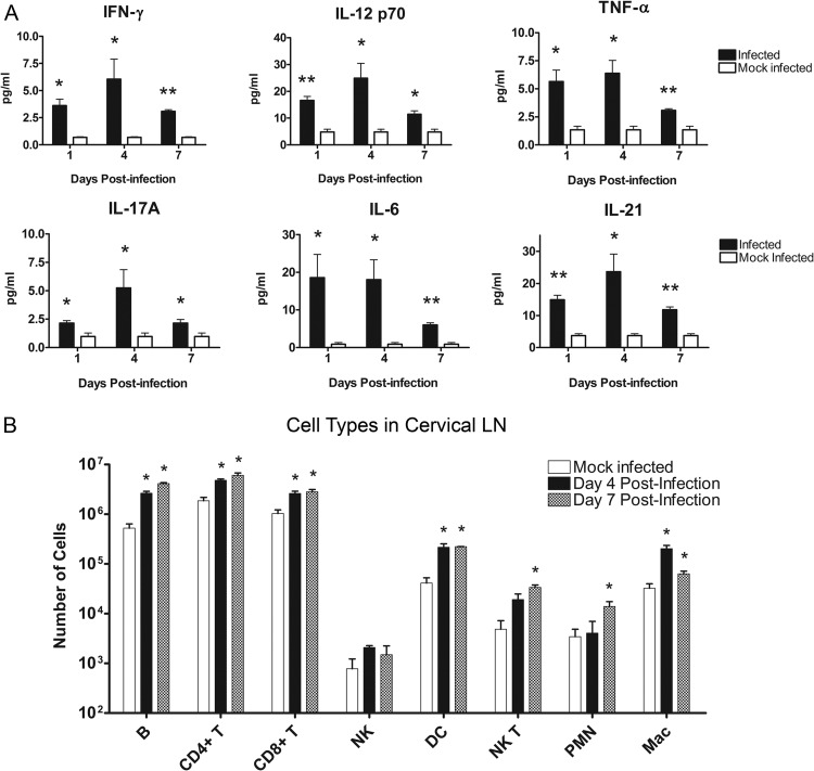
Fig 4.
Cytokine and immune cell profiles in the draining LN. Cervical LNs were removed from BALB/c mice after epicutaneous challenge with S. aureus-coated or mock infection with uncoated Morrow-Brown needles. (A) Supernatants from LN homogenates were analyzed for cytokines at days 1, 4, and 7 postinfection (n = 6 to 9 mice per group; experiments performed in duplicate; *, P < 0.05 compared to controls by Student's t test; **, P < 0.01 compared to controls by Student's t test). Bars represent SD. (B) Single-cell suspensions of LN cells at days 4 and 7 postinfection were prepared, and cell surface markers were stained with fluorescent antibodies. Samples were analyzed via flow cytometry (n = 3 mice per group; experiments performed in triplicate; *, P < 0.05 compared to controls by Student's t test). Bars represent SD.

