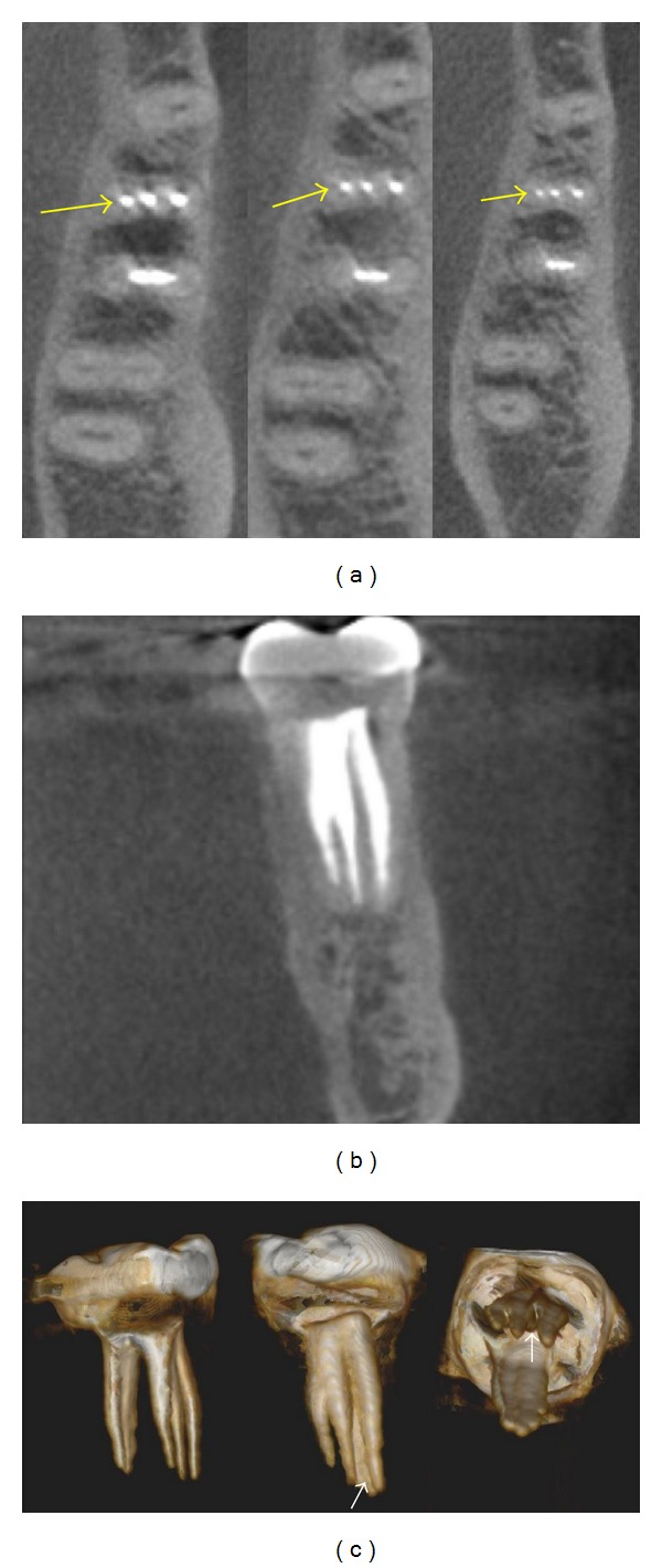
Figure 2.

(a) Axial CBCT slices on coronal, middle and apical root sections (arrows) showing three independent mesial canals. (b) Coronal CBCT view of mesial root with three filled root canals. (c). 3-Dimensional rebuild illustrating the internal configuration including a filled isthmus between the mesiobuccal and middle mesial canals (arrows).
