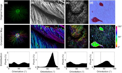
Fig. 5.

Fiber orientation detection via vector summation performed on (a) fluorescence microscopy image of DRG neurites, (b) scanning electron microscopy image of silk fibers, (c) second harmonic generation image of collagen in engineered bone, (d) picosirius red stained collagen fibers surrounding MCF10A cells (reproduced with permission24). For the orientation maps in the middle row, pixel-wise fiber angle measurements were color coded to the HSV color map in MATLAB, and these color maps were then multiplied by the grayscale intensity image to produce a final image.
