Figure 5.
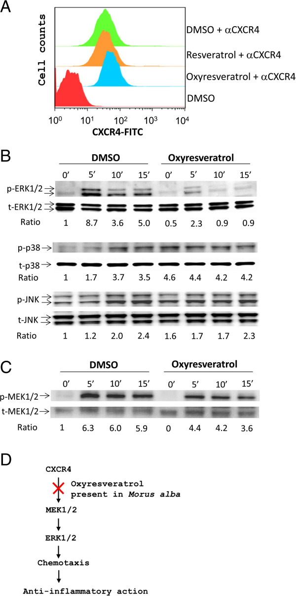
Effect of oxyresveratrol on CXCR4 signaling. (A) Jurkat cells were pre-treated with oxyresveratrol (2.5 μg/ml), resveratrol (2.5 μg/ml) or DMSO vehicle for 1 h at 37°C. After extensive washing, the cells were stained with αCXCR4 (1 μg/ml) and FITC-conjugated secondary antibody (1 μg/ml) or not. The expression level of CXCR4 on the cell membrane is shown in the histogram. Data are representative examples of 3 experiments. (B &C) The cells, pre-treated with oxyresveratrol or DMSO, were stimulated with SDF-1 (100 ng/ml) for 0 to 15 min. Cell lysates were analyzed by Western blot with primary antibodies (1 μg/ml) against MAPKs (B), MEK1/2 (C) or their phosphorylated forms (B &C) plus secondary antibodies (0.3 μg/ml). The ratio was obtained by normalizing the signal of the phosphorylated protein to that of the total protein. (D) A schematic model describing the mechanism by which oxyresveratrol present in M. alba can inhibit inflammation.

