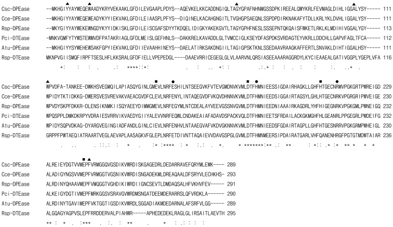
Figure 1. Multiple sequence alignment of DTEase family enzymes and their homologs.
Amino acid sequence for DPEase from C. scindens 35704 (Csc-DPEase; GeneBank accession No: CLOSCI_02528) was aligned with C. cellulolyticum H10 (Cce-DPEase; ACL75304), Ruminococcus sp. 5_1_39BFAA (Rsp-DPEase; ZP_04858451), P. cichorii DTEase (Pci-DTEase; BAA24429), A. tumefaciens DPEase (Atu-DPEase; AAL45544), and R. sphaeroides DTEase (Rsp-DTEase; ACO59490). The alignment was performed using ClustalW2 program (http://www.ebi.ac.uk/Tools/clustalw2/index.html). Amino acid residues that are identical in all the displayed sequences are marked by asterisks (*), strongly conserved or weakly conserved residues are indicated by colons (:) or dots (.), respectively. The symbol ▪, •, and ▴ represented the residues involved in the metal coordinating site, those responsible for the interaction between the enzyme and O1, O2, and O3 of D-fructose, and those providing a hydrophobic environment around the substrate around the O4, O5, and O6 of D-fructose, respectively (according to the crystal structures of C. cellulolyticum DPEase, A. tumefaciens DPEase, and P. cichorii DTEase).

