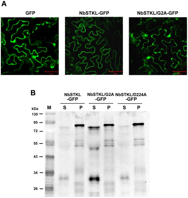
Figure 6. The subcellular localization of NbSTKL.
A. Subcellular localization of GFP, NbSTKL-GFP and NbSTKL/G2A-GFP was observed by confocal microscopy. Images were obtained by Olympus Fluoview FV1000 confocal microscope with 488 nm excitation. B. Western blotting was used to detect the localization of NbSTKL and its derivatives. The GFP fused NbSTKL (NbSTKL-GFP) and its derivatives NbSTKL/G2A-GFP and NbSTKL/D224A-GFP were transiently expressed by agro-infiltration onto N. benthamiana leaves. The total proteins were extracted and separated into the soluble fraction (S) and the pellet fraction (P). GFP antibody was used for Western blot assay. The scale bars represent 50 μm.

