Figure 3.
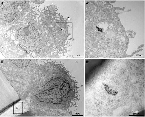
Particle uptake in the upper transwell cell layer. Ag-NPs were found in the upper cell layer of the transwell membrane (A) and in cells crossing the transwell insert (B) as aggregates inside vesicles at 24 h post-exposure (scale bar = 2 μm). Overall cell morphology upon Ag-NP exposure was similar to untreated control. A’ and B’ represent a higher magnification of the black marked box of the opposing picture (scale bar = 500 nm). B’ reveals particle agglomerates inside a multilamellar body.

