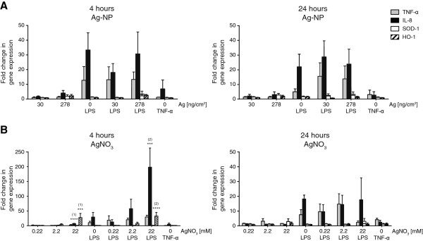
Figure 6.

Quantitative gene expression of pro-inflammatory and oxidative stress markers. Exposed cells were harvested 4 h and 24 h after exposure and mRNA levels of pro-inflammatory markers TNF-α (grey) and IL-8 (black) as well as oxidative stress markers SOD-1 (white) and HMOX-1 (striated) were analysed by real-time RT-PCR. Fold changes of gene expression compared to unexposed untreated controls were calculated with 2-ΔΔCt. Error bars represent the SEM for at least 3 independent experiments. A two-way ANOVA with a subsequent Bonferroni post-hoc test was performed. Values were considered significantly different compared to unexposed untreated control (1) and unexposed LPS treated control (2) with p < 0.001 (***) or p < 0.0001 (****).
