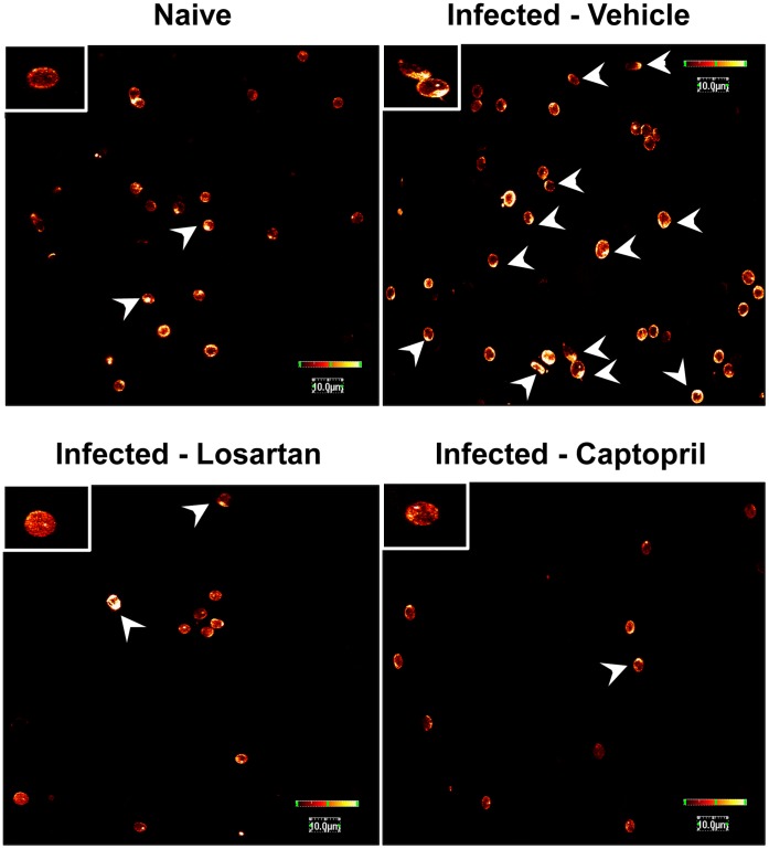
Figure 7. Ang II mediates F-actin polymerization in T lymphocytes during P.berghei ANKA infection on contact with endothelial basal membrane proteins.
Splenic T lymphocytes isolated from naive mice and mice infected with P. berghei ANKA treated with vehicle, losartan or captopril by gavage were stimulated for 1 h with fibronectin followed by F-actin cytoskeleton staining by phalloidin-Alexa Fluor 488. Cells were examined by confocal microscopy. Arrows indicate cells presenting spread morphology. The color bar on the right of the images represents the relative scale of fluorescence intensity.

