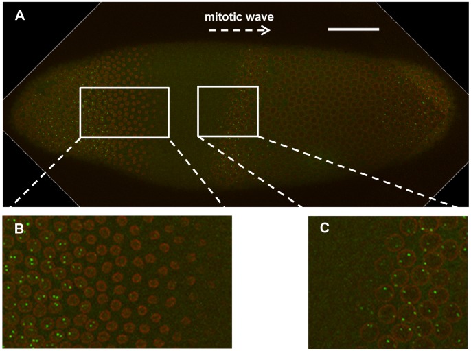
Figure 6. Active hb transcription at cycle 13 spans the entire interphase.
(A) Shown is a merged confocal image of an embryo undergoing mitosis. This embryo has the 13th mitotic wave on the anterior side, as evidenced by the breakdown of the nuclear envelope. Scale bar, 50 µm. (B) Shown is a higher magnification of a section of the image illustrating the reappearance of the cycle 14A nuclei and the hb intron dots (detected by an intronic probe) behind the mitotic wave. (C) Shown is a high magnification of a section of the image illustrating the disappearance of cycle 13 nuclear envelope and hb intron dots on the front of the mitotic wave.

