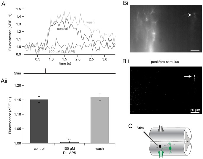
Figure 10. EIN-evoked Ca2+ signals are NMDA receptor-dependent.
A EIN stimulation evoked fluorescent Ca2+ signals in a motor neuron dendrite retrogradely filled with the Ca2+-sensitive dye, OGB1-dextran (5 mM), in the presence of NBQX (5 µM), glycine (1 µM), and strychnine (5 µM) in Mg2+-free ringer. Ca2+ signals were recorded before (black), during (dark grey) and after (light grey) the application of D/L-AP5 (100 µM). Pooled responses (Aii) demonstrate that D/L-AP5 significantly and reversibly abolishes NMDA receptor-induced changes in Ca2+ fluorescence. Error bars express ± SEM. B Image of the dendrite recorded in Ai. Localized EIN-evoked fluorescent change is shown in Bii (arrow, white). Scale bar = 20 µm. C Recording schematic: STIM = stimulating electrode, EIN = black, Ca2+-dye filled motor neuron = green.

