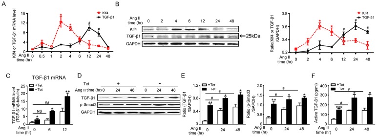
Figure 4. Klf4 controls Ang II-induced TGF-β1 production.
A, Klf4 and TGF-β1 mRNA levels were assessed by qPCR in Ang II-treated cardiac fibroblasts and were normalized to β-tubulin (n = 3). B, Klf4 and TGF-β1 protein levels were assessed by Western blot in Ang II-treated cardiac fibroblasts. GAPDH was the loading control (n = 3). Data are the mean ± SEM. *P<0.05, **P<0.01 vs. Ang II treatment for 0 hr. C-F, cardiac fibroblasts were co-infected with Ad-tTA and Ad-Klf4 and treated with Ang II. C, TGF-β1 mRNA levels were assessed by qPCR after Ang II treatment for 0, 6 or 12 hrs and normalized to β-tubulin (n = 4). D, TGF-β1 and phosphorylated Smad3 (p-Smad3) protein levels were assessed by Western blot after Ang II treatment for 0, 24 or 48 hrs. E, level of quantification of TGF-β1 and p-SMAD3 as a ratio of GAPDH in densitometric units was presented. n = 4. F, ELISA was used to analyze active TGF-β1 levels in the supernatant after Ang II treatment for 0, 24 or 48 hrs (n = 5). Data are the mean ± SEM. *P<0.05, **P<0.01 vs. control. #P<0.05, ##P<0.01 vs. control and Ang II treatment for 0 hr.

