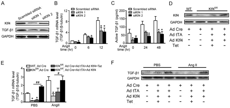
Figure 5. Ang II-induced TGF-β1 production is partly dependent on Klf4.
A–C, cardiac fibroblasts were transfected with siKlf4 and treated with Ang II. A, Western blot showed siRNA-mediated knockdown of Klf4 and TGF-β1 protein levels in cardiac fibroblasts. B, TGF-β1 mRNA levels were assessed by qPCR after Ang II treatment for 0, 6 or 12 hrs (n = 3). C, ELISA was used to analyze active TGF-β1 levels in the supernatant of Klf4- or scrambled-siRNA transfected cardiac fibroblasts after Ang II treatment for 0, 24 or 48 hrs (n = 3). *P<0.05 vs. scrambled siRNA. D–F, adult cardiac fibroblasts were isolated from mice carrying LoxP floxed Klf4 alleles. The cells were infected with Ad-Cre for Klf4 deletion, and infected WT cells were served as the negative control. Ad-Cre, Ad-tTA and Ad-Klf4 were co-infected to reintroduce Klf4 expression. D, 2 days after adenovirus infection, the cells were analyzed for Klf4 protein expression by Western blot. E, TGF-β1 mRNA levels were assessed by qPCR after Ang II treatment for 0 or 6 hrs in Klf4-deleted and Klf4-reintroduced fibroblasts and were normalized to β-tubulin (n = 3). Data are the mean ± SEM. *P<0.05 vs. Ad-Cre infected WT control; #P<0.05, ##P<0.01 vs. Ad-Cre infected Klf4-floxed fibroblasts. F, TGF-β1 protein levels were assessed by Western blot after Ang II treatment for 24 hrs.

