
Figure 7. Model for vegetal RNA localization.
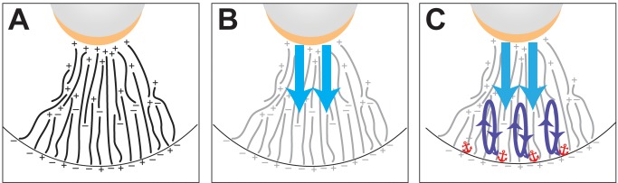
The vegetal cytoplasm is depicted, with the vegetal cortex at the bottom. The oocyte nucleus is shown in gray and the perinuclear cup is indicated in gold. (A) The oocyte microtubules are shown in black with orientation indicated by plus and minus. The proposed arrangement of microtubules is based on the appearance of a subpopulation of microtubule plus-ends at the vegetal cortex following breakdown of the mitochondrial cloud [12], which has been proposed to contain a microtubule organizing center [52]. (B) Vg1 mRNA enriched at the perinuclear cup is first transported by the dynein molecular motor in the upper vegetal cytoplasm in an initial highly directional step toward the vegetal cortex (blue). Microtubules are shown in grey. (C) Repeated cycles of bidirectional transport dependent on kinesin molecular motors occur in the lower vegetal cytoplasm (purple), until Vg1 mRNA exits the transport cycle by becoming anchored at the vegetal cortex (red).