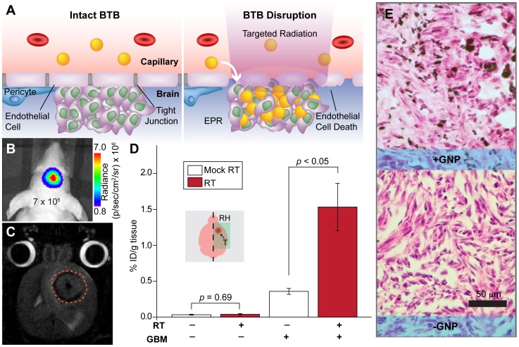
Figure 5. Radiation-induced modulation of the blood-brain barrier leads to increased uptake of GNPs in orthotopic GBM xenografts.
A. Schematic of BBB-disruption with targeted RT, which leads to endothelial cell death and loosening of tight junctions. B. Representative BLI image of a smaller, less disruptive GBM tumor (max BLI ∼106) used in this experiment. C. T2-weighted MRI image of a stereotactically implanted intracranial GBM tumor (approximated by dashed orange line). D. ICP-MS analysis of gold uptake in the right hemispheres of healthy brains and those with orthotopic GBM excised from mice 48 hours after i.v. injection of saline or 0.4 g Au/kg GNPs administered 7–14 days after 20 Gy RT or mock-irradiation. The right cerebral hemispheres of mice were orthotopically inoculated with 350,000 U251 cells. Tumors were allowed to grow until the measured BLI irradiance reached ∼106 p/sec/cm2/sr (approximately 2 weeks post-implantation), at which point the mice were given their respective treatments. Brains with tumors receiving RT (n = 4) prior to GNP administration show significant increase in EPR-driven gold accumulation compared to mock-irradiated controls (n = 4). Mock-irradiated (n = 3) and irradiated (n = 3) healthy brains, however, show no significant difference in gold uptake, suggesting that normal tissue may recover more quickly than tumor. E. Representative H/E staining of sections from orthotopic tumors with (+) and without (−) GNP injection.

