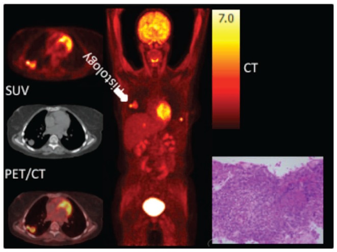
Figure 2.

A 28 year-old woman presented with asymptomatic pulmonary nodules associated with increasing EBV viral load in the peripheral blood four months following lung transplantation. Transaxial FDG-PET, CT and fused PET/CT images of the chest (left), maximum intensity projection (center) and histology of pulmonary nodule (right). FDG PET/CT showed a bilobar pulmonary nodule in the right lung (white arrow) and a lesion in the liver and left chest wall. Histological analysis of a core needle biopsy of the pulmonary nodule revealed an EBV-driven diffuse large CD20-positive lymphoid proliferation, compatible with a monomorphic PTLD, type DLBCL, NOS of non-germinal center B-cell origin. SUV: standardized uptake value.
