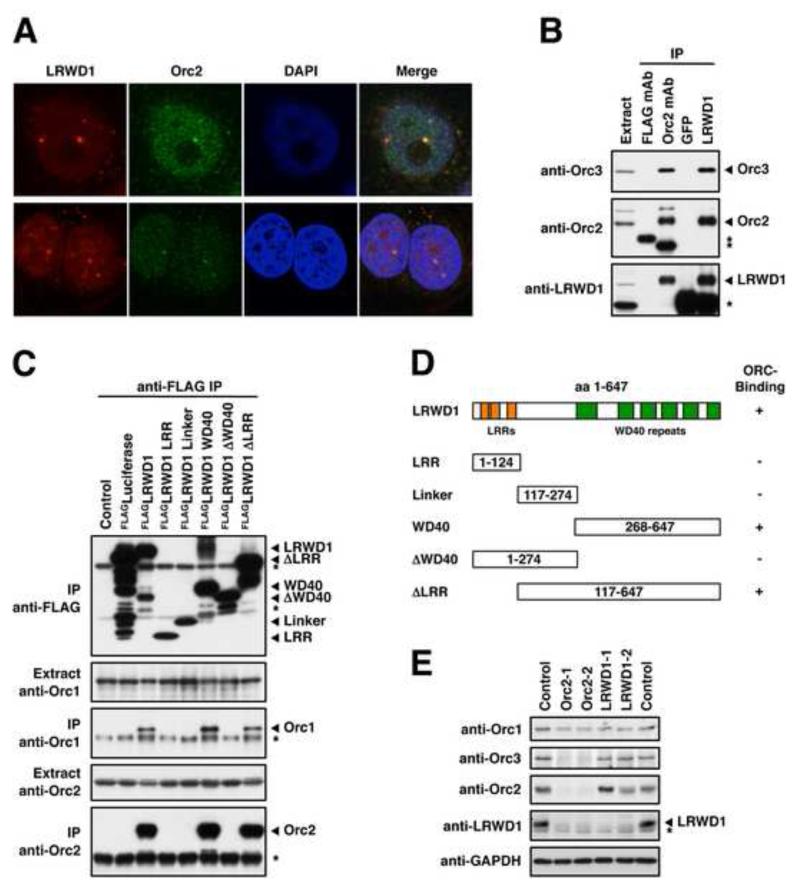
Figure 4. LRWD1 Interacts with the Origin Recognition Complex.
(A) LRWD1 co-localises with Orc2. IF staining of MCF7 cells with LWRD1 (2527) and Orc2 antibodies following pre-extraction shows co-localisation at distinct nuclear foci. (B) LRWD1 and ORC co-immunoprecipitate. LRWD1 and Orc2 were immunoprecipitated from MCF7 whole cell extracts and interacting proteins were detected by immunoblot as indicated. LRWD1 was immunoprecipitated using anti-LRWD1 (A301-867A) and detected using anti-LRWD1 (2527) antibodies. Anti-FLAG and anti-GFP antibodies were used as IgG negative controls. Asterisks mark bands derived from antibody heavy chains. (C) FLAG-tagged full length and truncated versions of LRWD1 were over-expressed in 293T cells and immunoprecipitated using an anti-FLAG antibody. 1 % of the input and 10% of the IP were separated by SDS-PAGE and Orc1, Orc2 and the FLAG fusions were detected by immunoblot. The asterisks mark bands derived from the anti-FLAG IP antibody. (D) Identities of the LRWD1 truncation constructs. Only deletions containing the WD40 repeats interact with ORC. (E) LRWD1 expression is Orc2-dependent. Expression levels of LRWD1 and ORC proteins in MCF7 cells were detected by immunoblot after transfection with siRNAs against LRWD1 and Orc2 as indicated. Cells were reverse-transfected twice, 56 h and 28h before harvesting. GAPDH serves as a loading control. The asterisk marks a cross-reactive band detected by the anti-LRWD1 (2527) antibody. See also Figure S3.

