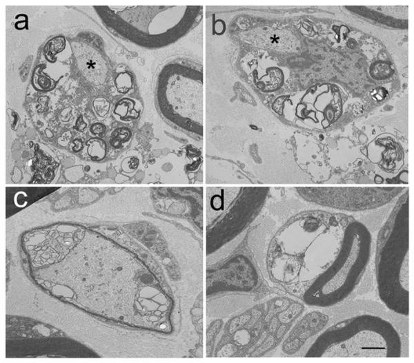
Fig. 3.

Electron micrographs of the ulnar nerve from an NPC affected cat confirmed the light microscopic findings. Myelin debris and vacuoles were noted in Schwann cell cytoplasm (a,b) with normal appearing axons (a,b asterisks) and Schwann cell nuclei (b). Inappropriately thin myelin sheaths (c) and large vacuoles containing lipid substrates (d) were noted. Bar in d = 0.87 μm for a and b, and 0.95 μm for c and d.
