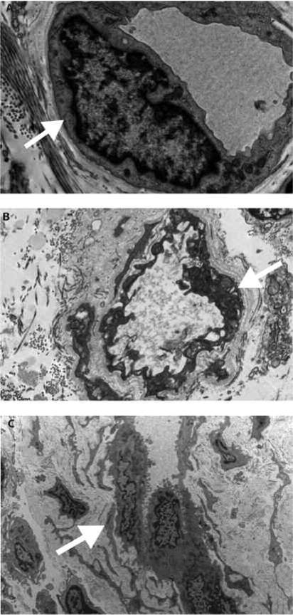
Figure 2.
Transmission electron microscopy. A. Control skin specimen shows a dermal capillary with thin basement membrane (arrow) and flattened endothelial cell. B. Capillary from a scleroderma case shows mild lamellation (arrow) of the basement membrane. C. A more extensive lamellation (arrow) of the basement membrane can be seen. The endothelial cells show abundant cytoplasm and enlarged nuclei.

