Figure 3.
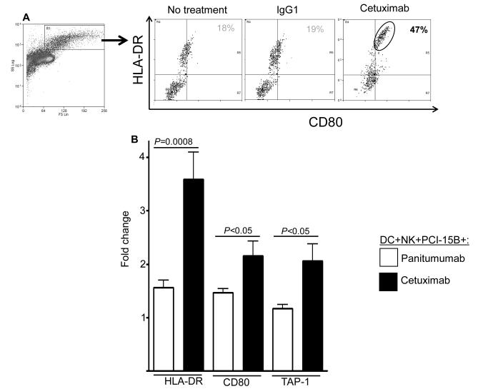
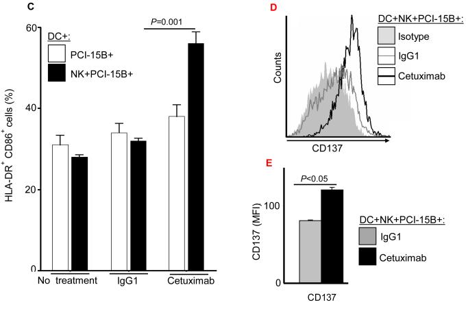
Enhancement of DC maturation by cetuximab-activated NK cells. (A) Histogram analysis of the upregulation of maturation markers HLA-DR+/CD80+ on DC co-cultured with NK:PCI-15B in the presence of cetuximab. (B) Analysis of upregulation of maturation markers on DC co-cultured with NK:PCI-15B in the presence of cetuximab or panitumumab. Expression level of HLA-DR (represented in MFI, n=13 donors), CD80 (represented in % positive cells, n=8 donors) and TAP-1 (represented in MFI, n=4 donors) on DC co-cultured with NK:PCI-15B (1:1:1 ratio) with no treatment or panitumumab or cetuximab (each 10μg/ml, 48h) were measured. The fold change of DC marker expression were related to levels on control DC co-cultured with NK:PCI-15B alone are shown. A two-tailed unpaired t-test was performed for statistical analysis. (C) Percentages of DC (HLA-DR+ CD86+) that were co-cultured with PCI-15B (1:1 ratio) or with NK:PCI-15B (1:1:1 ratio) with no treatment or with IgG1 or cetuximab (each at 10μg/ml, 48h) were measured. Data are representative of three experiments from three different donors. Enhancement of CD137 expression on DC by cetuximab-activated NK cells. (D-E) Histogram analysis of the upregulation of CD137 expression on DC co-cultured with NK:PCI-15B in the presence of IgG1 or cetuximab (each 10μg/ml, 48h). (B) The quantitative bar diagram showing the significant differences in CD137 expression is also shown. A two-tailed unpaired t-test was performed for statistical analysis.


