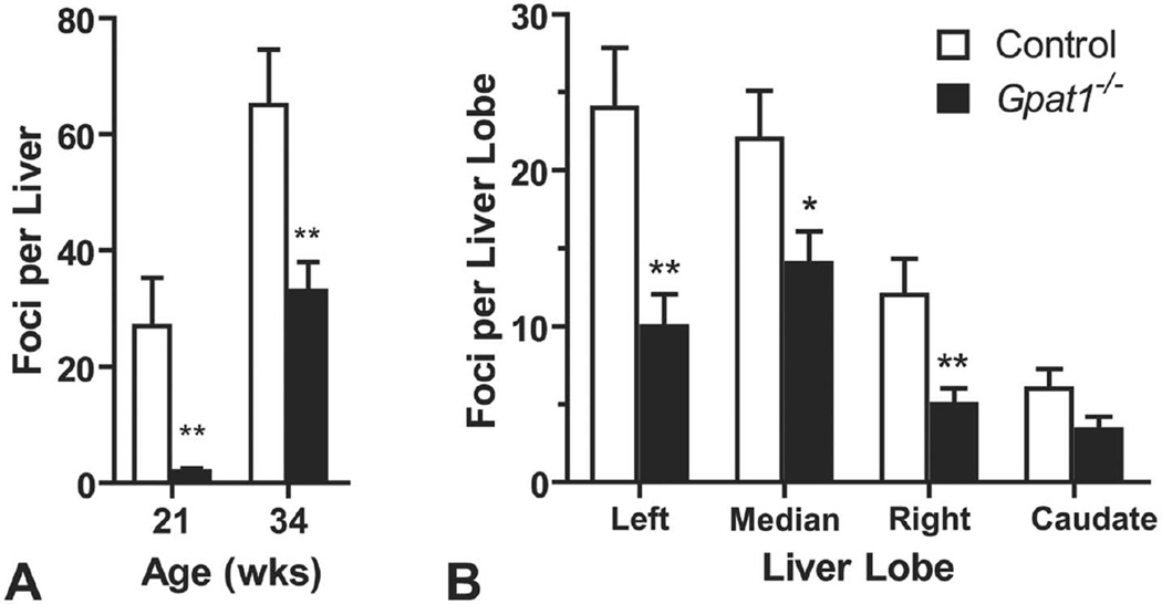
FIGURE 2.
Gpat1−/− mice had reduced susceptibility to liver cancer. (A) Average number of macroscopically visible liver nodules in control and Gpat1−/− 21-week-old mice (control, n = 12; Gpat1−/−, n = 11) and 34-week-old mice (control, n = 15; Gpat1−/−, n = 19). (B) Average of macroscopically visible nodules per liver lobe at 34 wk of age (n = 15). Data represent the average ± SEM; *p ≤ .05, **p ≤ .01, between genotypes.

