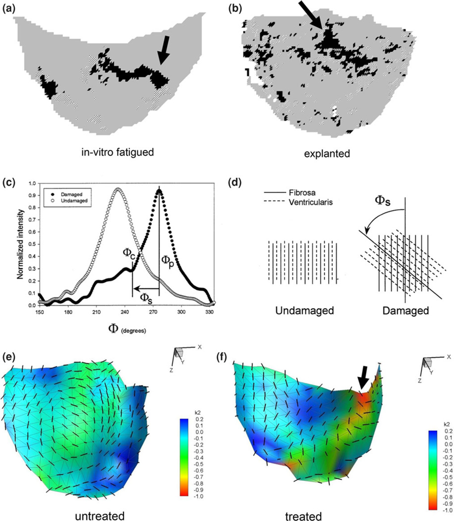
FIGURE 8.
Quantified collagen fiber disruption in porcine BHVs either (a) subjected to in vitro AWT28 or (b) explanted from patients after several years of in vivo function for non valve related problems.27 Here, regions of structural damage to the collagen fiber network are indicated as black or hatched areas (arrows).These damaged regions were detected by the presence of substantial skew, as shown in (c, d). Here, extracted collagen fiber distributions from structurally intact and damage regions show distinct increases in the skew, due to shearing of the fibers shown schematically in (d).17 The spatial locations of persistent bending in the treated leaflets compared untreated were located in similar regions of the leaflet (e, f). An arrow also shows regions of high curvature experienced by the leaflet.

