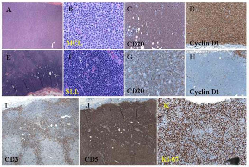
Figure 1.
Histologic and immunophenotypic features of a composite case of MCL and CLL/SLL. (A and B) Nodular pattern of atypical small lymphoid cells with a nodular pattern (H&E staining; original magnification: 20X and 100X). (C) Positive CD20 staining of atypical small lymphoid cells in both the internodular and nodular areas (immunoperoxidase staining; original magnification: 40X). (D) Cyclin D1 staining of atypical small lymphoid cells in the nodular area (immunoperoxidase staining; original magnification: 40X). (E and F) Atypical small lymphoid cells in the internodular area (H&E staining; original magnification: 20X and 100X). (G) Weak CD20 staining of atypical small lymphoid cells in the internodular area (immunoperoxidase staining; original magnification: 40X). (H) Negative cyclin D1 staining of atypical small lymphoid cells in the internodular area (immunoperoxidase staining; original magnification: 40X). (I) CD3 staining of normal small T-cells in the internodular area (immunoperoxidase staining; original magnification: 20X). (J) CD5 staining of atypical small lymphoid cells in both the internodular and nodular areas (immunoperoxidase staining; original magnification: 20X). (K) Ki-67 staining of a relatively high proliferation index in the nodular area (immunoperoxidase staining; original magnification: 40X).

