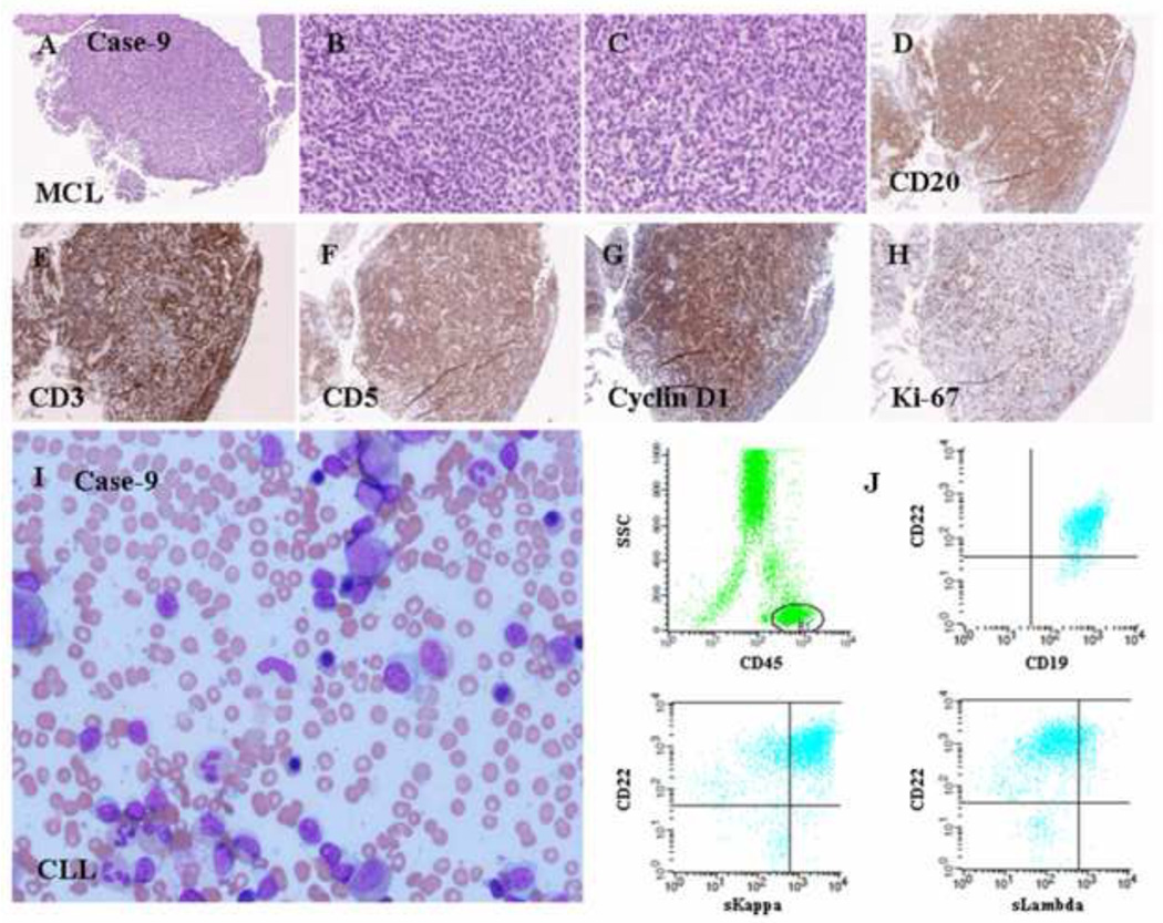
Figure 6.
Histologic and immunophenotypic features of a composite case of MCL and CLL/SLL. (A-C) A diffuse pattern of atypical lymphoid infiltration (H&E staining; original magnification: 20X and 100X). (D) CD20 staining of atypical lymphoid cells (immunoperoxidase staining; original magnification: 20X). (E) CD3 staining of normal small T-cells (immunoperoxidase staining; original magnification: 20X). (F) CD5 staining of atypical small lymphoid cells (immunoperoxidase staining; original magnification: 20X). (G) Cyclin D1 staining of atypical small lymphoid cells (immunoperoxidase staining; original magnification: 20X). (H) Ki-67 staining of a low proliferation index in the MCL area (immunoperoxidase staining; original magnification: 20X). (I) Bone marrow aspirate smear showing a few scattered atypical lymphoid cells with a clumped chromatin pattern. Flow cytometry revealed that these atypical lymphoid cells were positive for CD5, dim CD20, CD23, and monotypic kappa light chains, whereas cyclin D1 staining was negative based on the immunostaining of the bone marrow biopsy (data not shown).

