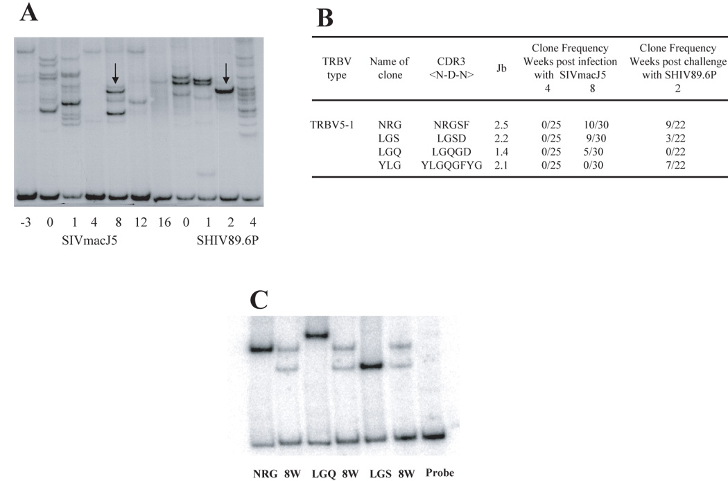
Figure 4. Analysis of the re-emergence of CD4 clonotypes in a protected animal.

A- TRBV5-1+ CD4 + clonotypes in macaque C94066F was analyzed by HTA from week −3 to week 4 post challenge. Arrows represent co-migrating bands at week 8 and week 2 post challenge. B- The PCR products from time points 4, 8 and 22 weeks (or 2 weeks post challenge) as shown by the arrows in figure A, were cloned and sequenced to identify their CDR3 regions as described in materials and methods. At week 8 two dominant CDR3 regions were identified, NRGSF with 10/25 clones and LGSD with 9/25 clones. At week 2 post challenge those two dominant TCR clones were detected. C- Co-migration of the DNA plasmids encoding the two dominant clones detected at week 8, NRG and LGS together with the PCR product from week 8. These results demonstrate that clones co-migrating at the same level are identical.
