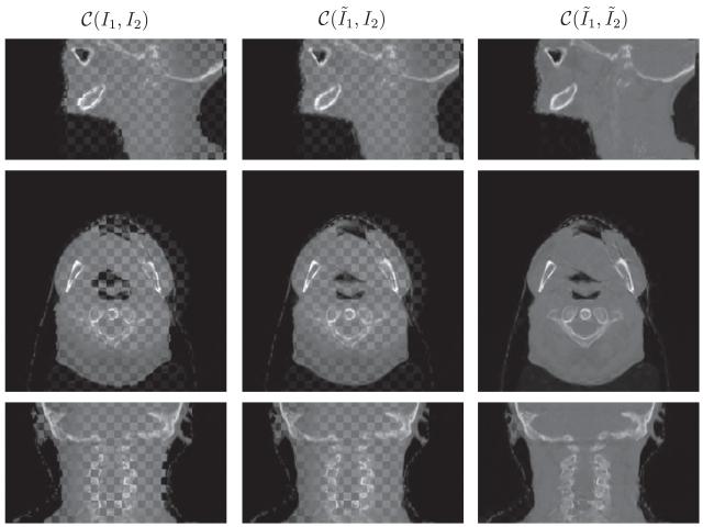
Fig. 7.
Performance on head-and-neck (B) in the checkerboard display. From top to bottom is the sagittal, axial and coronal view respectively. The results are resized in superior-interior direction corresponding to the physical space. Interpolation errors exists in the sagittal and coronal view, especially in the region of spine.

