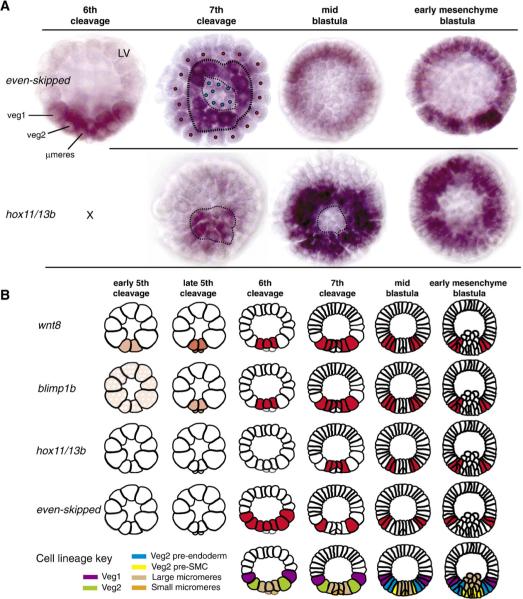
Fig. 7.
The relative expression patterns of wnt8, blimp1, hox11/13b, and eve. (A) Whole mount in situ hybridization (WMISH) of hox11/13b and eve, all panels vegetal view except where indicated lateral view, LV. At 6th cleavage, expression of eve is detectable in veg1, veg2, and micromeres. At 7th cleavage, the expression of eve wanes in veg1 and micromere descendents (red and blue dots, respectively), and eve is seen primarily in veg2 cells (second panel from left; dotted lines circumscribe the veg2 tier of cells). The loss of eve labeling in 7th cleavage micromere descendents corresponds with expression of hox11/13b in that territory at that stage (left panel; dotted lines mark micromere-veg2 boundary). At mid blastula stage, eve labeling is evident in presumptive endoderm cells, while hox11/13b is expressed in the presumptive mesodermal cells. By early mesenchyme blastula stage (right panels), eve expression has moved to veg1 endoderm, while the expression of hox11/13b is confined to veg2 descendents; n.b., the eve image is at a deeper plane of focus than the hox11/13b image. Expression of hox11/13b appears nested within the torus of eve expression. (B) Diagrammatic summary of WMISH results for wnt8, blimp1, eve and hox11/13b (this work, Smith et al., 2007, and Fig. S1). Lateral views are presented, which can be interpreted by the developmental key at bottom. Stippling in early 5th cleavage blimp1 figure indicates inferred maternal Blimp1 factor (Smith et al., 2007).

