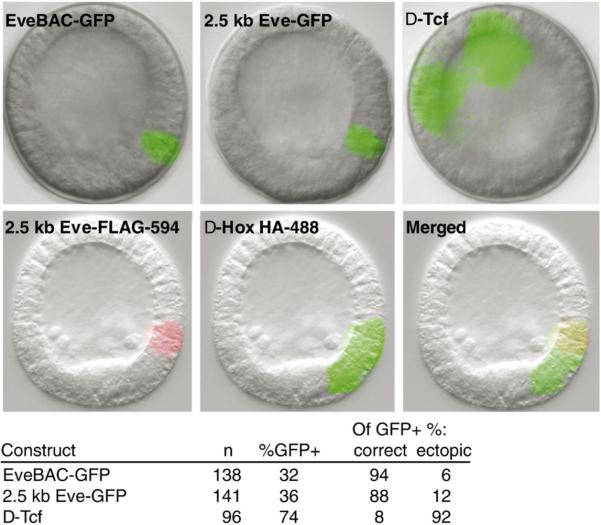
Fig. 9.
Effect of binding site disruption of even-skipped GFP reporters. (A) Representative fluorescent images of transgenic embryos harboring eve GFP reporter constructs. The top pair of panels display correct GFP reporter expression driven by the EveBAC-GFP recombinant and by a 2.5 kb Eve-GFP construct; expression is in veg1 endoderm cells in early mesenchyme blastula embryos. By contrast, embryos injected with the 2.5 kb Eve-GFP construct in which the Tcf site is mutated (Δ-Tcf; upper right panel) show gross ectopic expression in the ectoderm; not evident in these images is the relatively weak GFP reporter expression typical for this class of embryos. In the bottom three panels are experiments using the 2.5 kb Eve-GFP fragment bearing either FLAG or HA epitope tags. In these constructs, the Hox sites were, respectively, either left intact or disrupted. The normal (FLAG) construct is expressed the same as the parent 2.5 kb Eve-GFP construct (left panel); in contrast the Δ-Hox (HA) construct shows ectopic reporter expression in the veg2 clearance zone of endogenous eve expression (center panel). The lower right panel shows a merged image. (B) Table showing the quantitative effects of Tcf site disruption on eve GFP reporter activity. Loss of Tcf input leads to a greatly increased number of embryos displaying ectopic expression.

