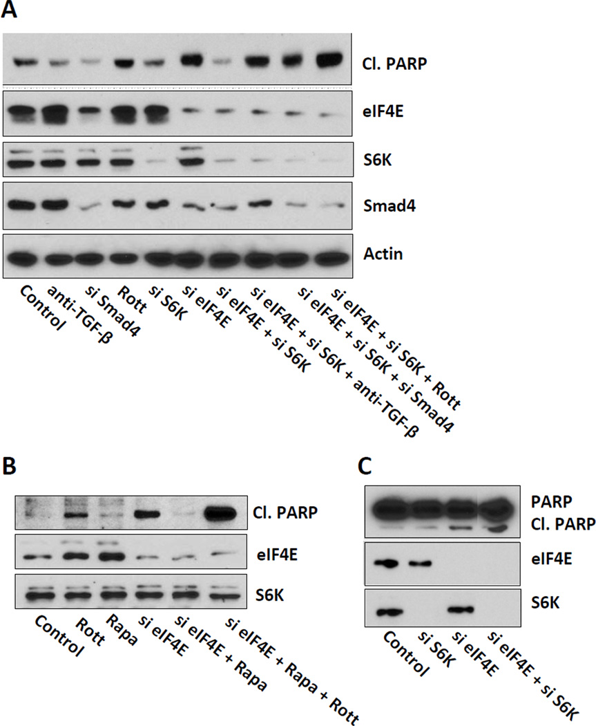
Figure 3.
Suppression of apoptosis by S6 kinase ablation is dependent on TGF-β signaling. (A) MDA-MB-231 cells were transfected at 50% confluence with negative control siRNA, siRNA for eIF4E, S6 kinase, or Smad4 as indicated. 24 hr later, the cells were put in fresh media with 10% serum and a neutralizing TGF-β antibody or rottlerin (Rott) (3 µM) where indicated. After another 18 hr, the level of cleaved PARP, eIF4E, S6K, Smad4, and actin by Western blot analysis. (B) MDA-MB-231 cells were transfected with negative control siRNA or siRNA for eIF4E as in (A). 24 hr later, the cells were put in fresh media with 10% serum and either 20 nM rapamycin or DMSO vehicle where indicated. Where indicated, rottlerin (Rott) (3 µM) was also provided. After another 18 hr, the level of cleaved PARP, eIF4E, and S6K by Western blot analysis. (C) BT-549 cells were prepared and transfected with negative control siRNA, siRNA for eIF4E, or S6 kinase as indicated. 24 hr later, the cells were put in fresh media with 10% serum and rapamycin (20 nM) where indicated. After another 18 hr, the level of cleaved PARP, eIF4E, and S6K by Western blot analysis. The data are representative of experiments repeated at least two times.

