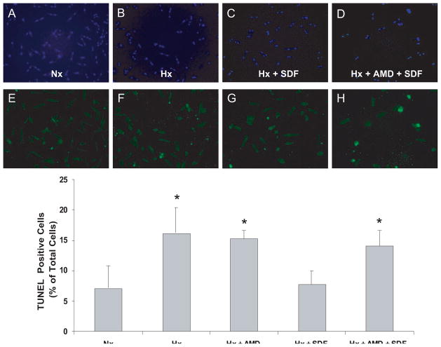
Figure 7.
SDF-1α inhibits apoptosis in cardiomyocytes. Myocytes pretreated with 40 μmol/L AMD3100 or vehicle for 40 minutes were stimulated with 25 nmol/L SDF-1α for 10 minutes followed by 1 hour of hypoxia (Hx) and 18 hours of reoxygenation. Apoptotic nuclei were determined by TUNEL assay. A and E, Normoxic (Nx) control; B and F, 1 hour of Hx and 18 hours of reoxygenation; C and G, 25 nmol/L SDF-1α before 1 hour of Hx and 18 hours of reoxygenation; D and H, 40 μmol/L AMD pretreatment with 25 nmol/L SDF-1α before 1 hour of Hx and 18 hours of reoxygenation; A through D, total nuclei (DAPI staining); E through H, TUNEL-positive myocyte nuclei (green fluorescent). Bottom, Summary of quantitative data from 3 independent experiments performed in duplicate. n = 3. *P<0.05 vs Nx.

