
Fig. 3.
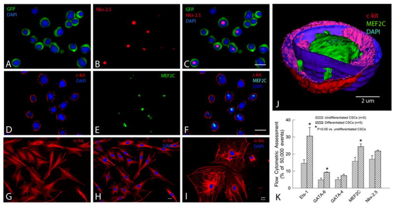
Profile and differentiation of murine CSCs. a–c Lin−/c-kit+/GFP+ CSCs cultured in growth medium at passage 4 express GFP in the cytoplasm (green) and, in some cases, the early cardiac transcription factor Nkx-2.5 in the nucleus (red). d–f: Lin−/c-kit+/GFP+ CSCs cultured in growth medium at passage 2 express c-kit on the membrane (red) and, in some cases, the early cardiac transcription factor MEF2C in the nucleus (green). CSCs cultured in differentiation medium in Matrigel for 10 days (g–h) or in the culture plate without Matrigel for 50 days (i) express the cardiac marker α-sarcomeric actin in the cytoplasm (red) (g–i) and display a cardiac myocyte-like morphology (i). j A 3-D computer reconstruction based on 42 confocal microscopic images showing a CSC that expresses c-kit (red) on the membrane and the early cardiac transcription factor MEF2C (green) in the nucleus. The nuclear membrane is stained with DAPI (blue). k FACS analysis of CSCs cultured for 10 days in differentiation medium shows increased expression of the lineage markers Ets-1 (endothelial), GATA-6 (smooth muscle), and GATA-4, MEF2C, and Nkx-2.5 (cardiac), demonstrating that lin−/c-kit+/GFP+ CSCs are heterogeneous and possess the potential to commit to all three components of the cardiac lineage. Data are mean ± SEM. Bars 10 μm