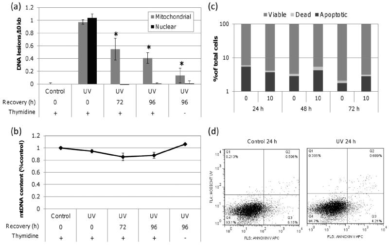
Figure 1. UVC-induced mtDNA damage is removed slowly.
(a) Significant mtDNA damage removal was observed in cells dosed with 10 J/m2 UVC by 72 h compared to initial lesion frequency (one-way ANOVA, effect of time point P = 0.0140; Fisher’s PLSD, P = 0.0316 for 72 h and P = 0.0047 for 96 h). nDNA damage was repaired to baseline by 72 h. (b) mtDNA content was comparable to control throughout the recovery period. (c) No significant decrease in cell viability was observed in cells exposed to 10 J/m2 as measured by Annexin V APC and Hoechst 33258. (d) Representative control and UVC dot plots at 24 h post exposure as measured by FACS with viable (Q4), apoptotic (Q3) and dead (Q2) cell frequencies defined. Bars ± s.e.m.

