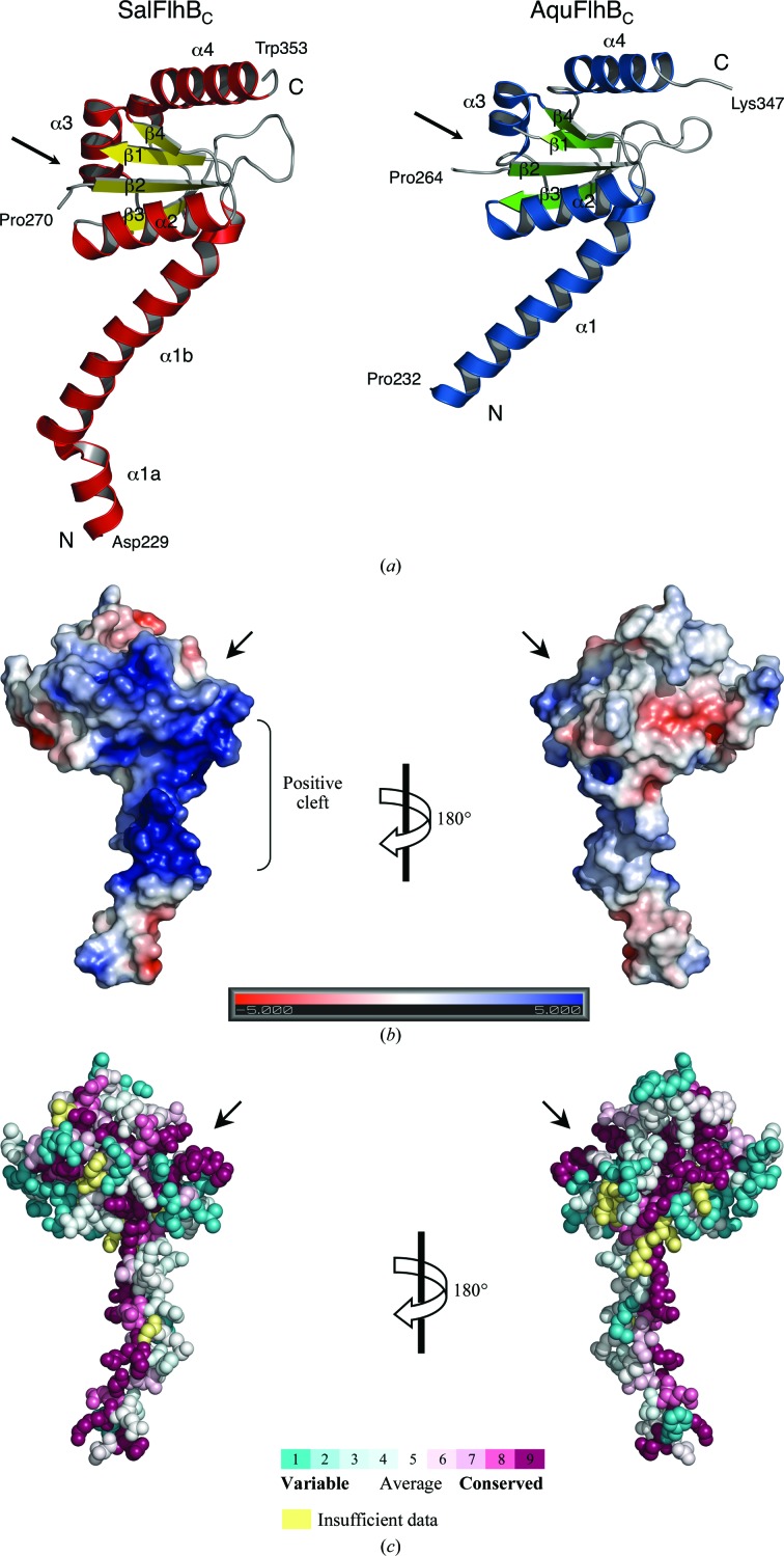
Figure 2.
Structure of flagellar FlhBc. (a) Ribbon representation of the crystal structures of Salmonella and Aquifex FlhBC. (b) Electrostatic potential mapped onto the surface of Salmonella FlhBC. Electrostatics were calculated using the APBS software (Baker et al., 2001 ▶) and plotted at ±5kT e−1. (c) Evolutionarily conserved residues of FlhBC. The figure was prepared with ConSurf (http://consurf.tau.ac.il/; Ashkenazy et al., 2010 ▶). Residues are coloured according to the conservation in amino-acid sequences of 200 different FlhB proteins. Arrows mark the position of the autocleavage site between β1 and β2.

