Figure 1.

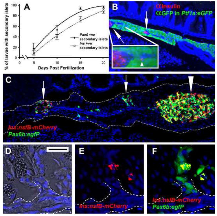
Secondary islets are established during the first 3 weeks of larval life and are associated with the main pancreatic duct. (A) The results of counting, over time, the occurrence of secondary islets in either Tg(P0-pax6:GFP)ulg515 or Tg(ins:mCherry)jh2 larvae. Starting at 5 days, few larvae display a secondary islet as detected by either transgene. By 20 days almost all larvae have at least one secondary islet.
(B) Paraffin section through the pancreas of a 28dpf, Tg(ptf1a:eGFP)jh1 juvenile following immunofluorescent detection for GFP (green) and insulin (red). The main pancreatic duct can be distinguished as a GFP negative strip of cells running through the middle of the pancreatic tail. Two secondary islets can be seen in the pancreatic tail (↑) closely associated with the duct. Inset is an enlargement of the secondary islet on the left. An isolated β-cell can be seen located right on the duct (◄). (C) Cryosection of adult pancreas lobe from a Tg(ins:nfsB-mCherry)jh5;Tg(P0-pax6b:GFP)ulg515, which includes the principal islet (large arrowhead) and two significant secondary islets (↑). (D,E,F) Close up of secondary islet (second from the left); (D) bright field image merged with nuclei staining reveals a morphological epithelial duct (outlined by white dotted line). (E) β-cells can be discerned by red fluorescence and are located in close association of an offshoot of the duct. (F) Pax6b expressing cells are marked green and in a merged image demonstrate endocrine cells adjacent to the duct. (D,E,F scale bar = 20μm)
