Figure 1.
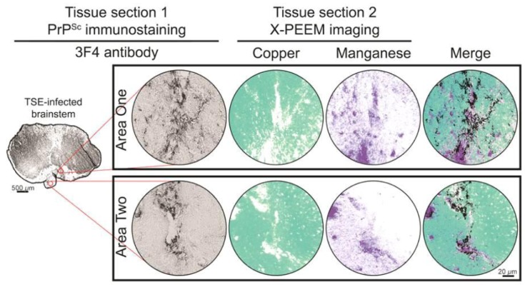
A coronal section (Tissue section 1) of a brainstem from a HY TSE-infected hamster was immunostained for PrPSc with monoclonal antibody 3F4. Areas with PrPSc deposits (as indicated by a black reaction product) larger than 7 µm were identified for analysis by X-PEEM on Tissue section 2, a directly adjacent tissue section. In Tissue section 2, copper and manganese distributions were assessed by X-PEEM in each area of PrPSc deposition. The presence or absence of copper is indicated by aqua pseudocoloring or white, respectively. Purple pseudocoloring or white represents the presence or absence of manganese, respectively. Superimposing PrPSc immunostaining micrographs with copper and manganese images (Merge) indicates the spatial distribution of all signals. Scale bars are labeled with the appropriate lengths for the corresponding images.

