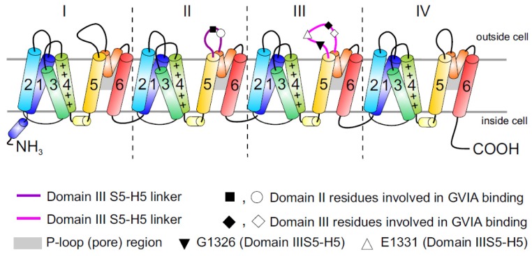
Figure 1.
Topology of Cav channels: Represented is the pore-forming α1 subunit of the Cav2.2 channels. This large protein consists of four homologous transmembrane domains (I–IV) and each domain contains six segments (S1–S6) and a membrane-associated P loop between S5 and S6 (represented in orange/grey) where the binding site of ω-conotoxins is localized. Circles, triangles and rectangles represent the localization of specific residues described to be important for binding of Cav2.2 to the ω-conotoxin GVIA [11].

