
Fig 5. Papillary urothelial carcinoma, high grade, non-invasive.
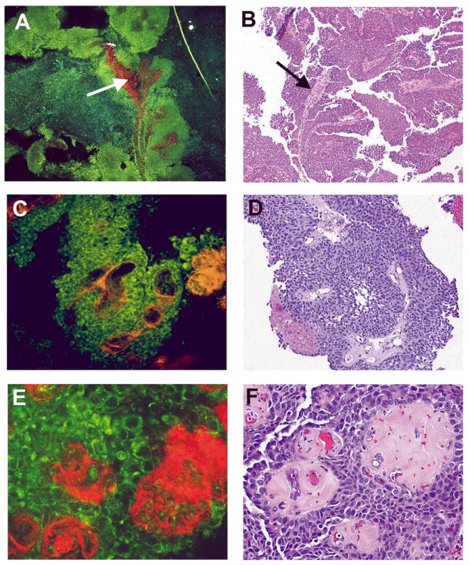
Multiphoton microscopy (MPM) image (A) and corresponding H&E image (B) at low magnification showing the papillary nature of the lesion (complex, thickened papillae with thin fibrovascular cores; arrow). MPM image (C) and corresponding H&E image (D) at high magnification showing thickened fibrovascular cores and disorderly arrangement of cells. MPM image (E) and corresponding H&E image (F) at high magnification showing cells with marked pleomorphism, high Nuclear:Cytoplasmic (N:C) ratio, and hyalinized fibrovascular cores. MPM magnifications: A = 48X, C = 240X, E = 480X; H&E magnifications: B=40X, D=200X, F=400X).