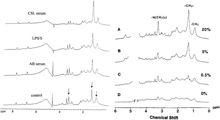
Figure 4.
MR-visible lipid spectra can be modulated by external serum levels. On the left, 1H MR 1D spectra obtained from neutrophils incubated in Hanks' balanced salt solution (control), AB serum containing low levels of fatty acids, lipopolysaccharide (LPS), and CSL serum containing high levels of fatty acids. The levels of lipid accumulation are higher in the CSL serum than in cells stimulated with LPS. The arrows indicate the lipid methylene peak at 1.3 ppm, the methyl peak at 0.9 ppm, and the taurine/choline peak at 3.2 ppm. The internal standard, p-aminobenzoic acid produces two doublets, one at 6.83 and the other at 7.83 ppm. On the right hand side, the level of mobile lipids in human mixed peripheral blood lymphocyte cultures is directly dependent on the amount of human serum in the culture medium. Note in both sets of spectra the presence of resonances at 3.2 and 3.4 ppm indicating high levels of taurine as well as choline. Taurine is present in the cytoplasm of immune cells at high concentrations where it acts as an osmolyte and radioprotectant.

