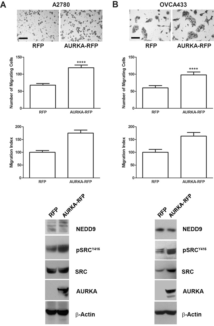
Figure 3. Enforced expression of AURKA stimulates ovarian carcinoma cell migration.
A2780 (A) and OVCA433 (B) cells were transiently transfected with an RFP expression construct or an AURKA-RFP expression construct for 24 h and then subjected to migration assays and immunoblot analyses. Bright-field images (10×) of migrating cells from representative inserts are shown (scale bar = 100 µm). Quantification of migration for A2780 (A) and OVCA433 (B) is depicted in the top graphs as the mean number of migrating cells ± SE (n = 3). Bars labeled with asterisks are statistically significant (****P < 0.0001) as analyzed by the Mann-Whitney test. The migration index (relative to vehicle-treated control) is depicted in the bottom graphs as the mean relative to RFP-transfected cells ± SE. Total proteins from cells transfected with RFP and AURKA-RFP were also immunoblotted with antibodies against NEDD9, pSRCY416, total SRC, AURKA, and β-actin (loading control).

