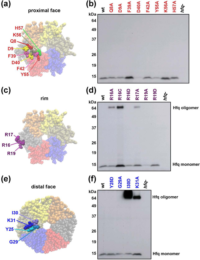
Fig. 1.
Chromosomal Hfq mutants. (a, c, e) Space-filling representation of the E. coli Hfq crystal structure (PDB 1HK9) showing the locations of amino acids mutated viewed from the proximal face (Q8, D9, F39, F42, K56 and H57), the rim (R16, R17 and R19), or distal face (Y25, G29, I30 and K31). (b, d, f) Hfq protein levels in mutant strains. Extracts were prepared from derivatives of SG30200 (PBAD-rpoS-lacZ) carrying wild-type and mutant hfq alleles (see Table S5); cells were grown in LB medium at 37 °C to early stationary phase (OD600 ~ 1.0). The levels of Hfq protein were determined by immunoblot analysis using anti-Hfq serum and ECL Western Blotting System. Ponceau S staining of the immunoblots showed that equal total protein amounts were loaded.

