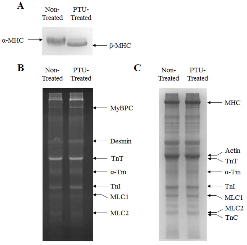
Figure 2.

Effect of five weeks of PTU treatment on isoform expression of MHC and other myofilament proteins, and on myofilament protein phosphorylation status in mouse ventricular preparations. Separation of α-MHC and β-MHC isoforms was done on 6.5% SDS polyacrylamide gels, as shown in panel A. Separation of other myofilament proteins was done on 12.5% gels. After separation, protein phosphorylation of myofilament proteins was visualized by UV-transilluminescence following Pro-Q diamond staining of 12.5% gels, as shown in panel B. After Pro-Q imaging, gels were stained with coomassie blue to visualize whether the isoform expression profiles of proteins other than MHC were altered by PTU treatment, as shown in panel C.
