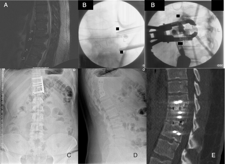
Fig. 2.

Twenty-four-year-old male with myelopathy. a Sagittal T2 MRI demonstrating disc herniations at T11-12 and T12-L1 causing cord compression. b Intraoperative fluoroscopic images demonstrating retractor positioning and localization of the interspace. c, d AP and lateral radiographs demonstrating Nuvasive PEEK cages with a lateral thoracic spine locking plate. e Sagittal CT at 8 months after the index surgery shows completed fusion of the instrumented levels.
