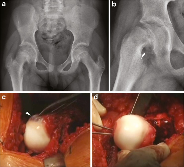
Fig. 1.
Acute on chronic slipped capital femoral epiphysis. a A decreased epiphyseal height and a break in Klein’s line is noted in the right hip on the AP pelvic radiograph. b Frog-leg lateral reveals callus formation at the posterior femoral neck with a severe displacement of the epiphysis (arrow). c Intraoperative image of remodeling of the anterolateral femoral head–neck junction (arrowhead). d Reduction and antegrade fixation of the epiphyseal fragment through the fovea centralis.

