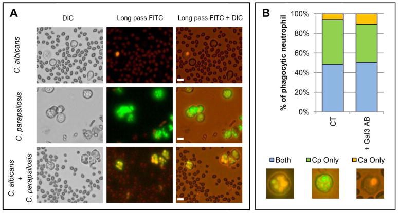
Figure 7. Neutrophils do not selectively phagocytose C. parapsilosis yeast over C. albicans yeast during coincubation.
All images are representative fields of at least three different donors. Bar = 10 microns. (A) Images of neutrophil phagocytosis of C. albicans dyed orange with ethidium bromide alone (top panel), C. parapsilosis dyed green with FITC alone (middle panel), or both orange C. albicans and green C. parapsilosis together (bottom panel). (B) Percent of total phagocytic neutrophils containing only C. albicans (Ca) or C. parapsilosis (Cp) or both species together. Images under the graph represent neutrophils containing both C. albicans and C. parapsilosis (left panel), green C. parapsilosis only (middle panel), or orange C. albicans only (right panel). Phagocytic contents of untreated neutrophils (CT) and neutrophils pretreated with gal3 blocking antibody (+Gal3 AB) were evaluated.

