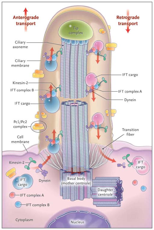
Figure 1. Structure of the Cilium and Intraflagellar Transport.
The cilium is a hairlike structure on the cell surface that consists of a microtubule-based axoneme covered by a specialized plasma membrane, which is assembled from the basal body, or mother centriole. Transition fibers act as a filter for molecules passing into or out of the cilium. Nephrocystin-1 is localized at the transition zone of epithelial cells (not shown).1 Axonemal and membrane components are transported in raft macromolecular particles (complexes A and B) by means of intraflagellar transport (IFT) along the axonemal doublet microtubules2 toward the tip complex by heterotrimeric kinesin-2. Mutations of Kif3a cause renal cysts and aplasia of the cerebellar vermis in mice.3 Retrograde transport occurs by means of the motor protein cytoplasmic dynein. (Adapted from Bisgrove and Yost.4)

