Fig. 2.
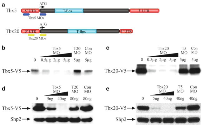
An example of translation-blocking MOs targeting the 5′UTR region of X. laevis genes Tbx5 and Tbx20. (a) Schematics showing the MO target locations on Tbx5 and Tbx20 mRNA. (b–e) Western blot analysis of Tbx5 and Tbx20 translation inhibition by specific MOs using V5 antibody. (b) Tbx5 MOs inhibit the in vitro translation of Tbx5 RNA fused with V5 epitope in a dose-dependent manner. Both Tbx20 (T20) MO and Con MO are unable to inhibit Tbx5-V5 translation. (c) Tbx20 MOs inhibit the in vitro translation of Tbx20 RNA tagged with V5 epitope in a dose-dependent manner. Both Tbx5 (T5) MO and Con MO cannot inhibit Tbx20-V5 translation. (d) Embryos were injected with 2 ng Tbx5-V5 RNA and Tbx5 MOs. Translation of Tbx5 was inhibited in vivo by Tbx5 MO in X. laevis animal caps in a dose-dependent manner as assessed by anti-V5 western blot. Tbx20 and Con MO are unable to reduce Tbx5-V5 protein expression. The membrane was reprobed with anti-Shp2 antibody as a loading control. (e) Translation of 2 ng Tbx20-V5 RNA is inhibited by Tbx20 MOs in X. laevis animal caps in a dose-dependent manner. Tbx5 and Con MO are unable to reduce Tbx20-V5 protein expression, assayed by western blot with anti-V5 antibody. Reproduced from (24) with permission from The Company of Biologists.

